THE VIRTUAL EDGE: Lab 3 Bacterial Staining Techniques II |
|||||||||||||||||||||||||||||
Gram Stain: Background & IntroductionGram +Gram-positive cell walls have a thick peptidoglycan layer beyond the plasma membrane. Characteristic polymers called teichoic and lipoteichoic acids stick out above the peptidoglycan and it is because of their negative charge that the cell wall is overall negative. These acids are also very important in the body’s ability to recognize foreign bacteria. Gram-positive cell walls stain blue/purple with the Gram stain.
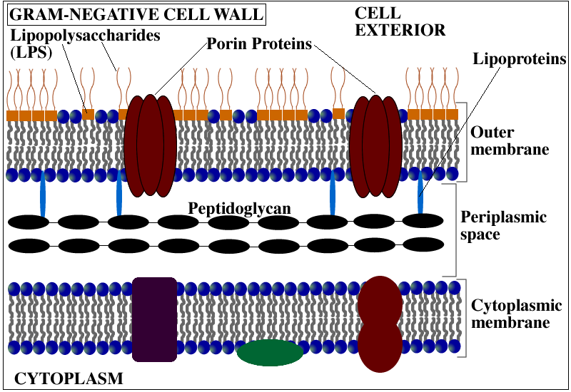
Gram -Gram-negative cell walls are more complex. They have a thin peptidoglycan layer and an outer membrane (lipopolysaccharide) beyond the plasma membrane. The space between the plasma membrane and the outer membrane is called the periplasm. The outer leaflet of the outer membrane is composed mainly of a molecule called lipopolysaccharide (LPS). LPS is an endotoxin that is important in triggering the body’s immune response. Gram-negative cells will stain pink with the Gram stain.
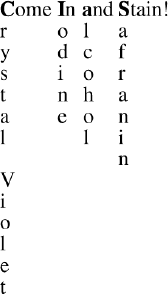
An easy way to remember the steps of the Gram stain is...
|
|||||||||||||||||||||||||||||
| Lab 3 / Gram Stain / Acid Fast Stain / Lab 3 Organisms Please take a few minutes to fill out a brief survey about your experience using the Virtual Edge: https://docs.google.com/forms/d/1yGbkF0KM92WBSk-IgS-EkjxkTKTQwhzuXmDsVpwRDoU/viewform Please email comments/problems to cboggs@uwyo.edu |
|||||||||||||||||||||||||||||
Rachel Watson, M.S. |
|
||||||||||||||||||||||||||||


The Virtual Edge by http://www.uwyo.edu/virtual_edge/ is licensed under a Creative Commons Attribution-Noncommercial-Share Alike 3.0 United States License