THE VIRTUAL EDGE: Lab 4 Bacterial Staining Techniques III |
|
Endospore Stain
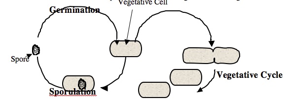
The most important endospore-forming bacteria are members of the genera Bacillus and Clostridium, both of which are Gram-positive rods. An endospore is a dense, multilayered structure that contains the genetic material of the bacterial cell. Endospores are formed within a vegetative bacterial cell when the environmental conditions no longer support cell growth. As the vegetative cell dies, the endospore is released into the environment where it can survive indefinitely in the presence of many environmental stresses, such as dessication, extremes in temperature, radiation, and lack of nutrients. When more favorable conditions arise the endospore germinates, again forming a viable vegetative cell. The presence of endospores in a bacterial culture can be detected by staining with malachite green. Because the endospore coat is so tough, steam is used to enable dye penetration. After washing, only the endospores will retain the primary stain Malachite green. Safranin is then used as a counterstain for vegetative cells. The endospore stain is a differential stain because it differentiates spore-formers from non spore-formers. Note: Formation of an endospore. The spore stains green and the vegetative cells stain red to orange. |
|
| Lab 4 / Endospore Stain / Capsule Stain / Lab 4 Organisms Please take a few minutes to fill out a brief survey about your experience using the Virtual Edge: https://uwacadweb.uwyo.edu/MOLB_SURVEY/ Please email comments/problems to cboggs@uwyo.edu |
|
Rachel Watson, M.S. |
|

The Virtual Edge by http://www.uwyo.edu/virtual_edge/ is licensed under a Creative Commons Attribution-Noncommercial-Share Alike 3.0 United States License