THE VIRTUAL EDGE: Lab 2 Bacterial Staining Techniques I
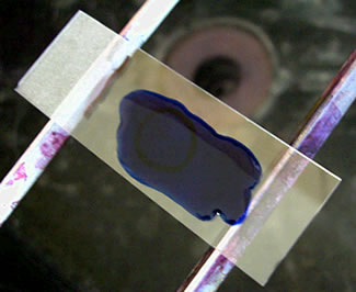
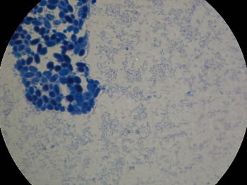
Direct Stain:

The cell wall of most bacteria has an overall net negative charge and thus can be stained directly with a single basic (positively charged) stain or dye. This type of stain allows us to observe the shape, size and arrangement of bacteria.

  1. Use the smear prepared in the previous procedure. Staining is done at the sink.
  2. Add several drops of Methylene blue, enough to cover the smear, and wait 1 min.
    Methylene Blue
  3. Rinse the slide with water from the squirt bottle.
    Rinse
  4. Blot the slide with bibulous paper. Redraw the focus line on the top of the slide if necessary.
    Blot
  5. Focus on the line with the 10X objective. Once you have focused on the specimen using the 10X objective, move the 40X objective lens into position. Use the fine adjustment knob to bring the specimen into focus. Now use the oil immersion procedure to view the specimen using the 100X:
    Specimen
    Note: Saccharomyces cerevisiae is a species of yeast. It is a relatively large single-celled eucaryotic organism. Escherichia coli is a "tiny" rod shaped bacteria (prokaryotic).
  6. Draw the organisms observed in the microscopic field.

Lab 2 / Smear Preparation / Direct Stain / Negative Stain / Oil Immersion Procedure / Terms & Definitions / Lab 2 Organisms

Please take a few minutes to fill out a brief survey about your experience using the Virtual Edge: https://docs.google.com/forms/d/1yGbkF0KM92WBSk-IgS-EkjxkTKTQwhzuXmDsVpwRDoU/viewform

Please email comments/problems to cboggs@uwyo.edu


Rachel Watson, M.S.
AG 5010
766-3524
Cell: 307-760-2942
rwatson@uwyo.edu

Microbiology Lecture WebsiteMicrobiology Lab WebsiteThe Virtual Edge Homepage

Creative Commons License
The Virtual Edge by http://www.uwyo.edu/virtual_edge/ is licensed under a Creative Commons Attribution-Noncommercial-Share Alike 3.0 United States License.

Creative Commons License
The Virtual Edge by http://www.uwyo.edu/virtual_edge/ is licensed under a Creative Commons Attribution-Noncommercial-Share Alike 3.0 United States License