THE VIRTUAL EDGE: Lab 2 Bacterial Staining Techniques I
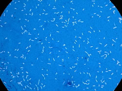
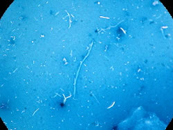
Negative Stain

In contrast to direct stains that bind to bacteria directly, a negative stain colors the background of a smear rather than the bacteria. These stains have negatively charged functional groups so they can't bind directly to negatively charged bacteria. The advantages of negative staining are:

  1. bacteria are not heat fixed so they don't shrink, and
  2. some bacterial species resist basic stains (Mycobacterium) and one way they can be visualized is with the negative stain.

However, negative staining does not differentiate bacteria, one can only determine morphology.

Procedure:

  1. Using a flamed inoculating loop, place 2-3 loopfuls of Congo Red in two separate circles on a clean slide. There is no need to add water to the Congo Red.
    Congo Red
  2. Using a flamed inoculating NEEDLE, pick up a small amount of Bacillus subtilis and stir it into one drop of Congo Red.

  3. Use a toothpick to scrape material from your teeth near the gumline and stir this into the second drop of Congo Red. Be sure to keep the two drops separate.
    Use Toothpick
  4. Air dry - DO NOT HEAT FIX.
  5. Flood the slide with acid-alcohol (95% ethanol, 3% HCl) until it turns blue. This generally takes ~ 2 seconds. Drain the excess acid-alcohol but do not wash the slide.
    Flood with acid-alcohol
  6. Allow the slide to air dry; do not blot.
  7. Examine both slides. First focus using the 10X objective. You will not be able to see individual organisms, but you should be able to focus on the stain. Then move to 40X and finally to the oil immersion lens with oil.
    b. SubtilisTooth Scraping
    Note: Organisms appear white (colorless) against a blue stained background.
  8. Draw a typical microscopic field for each slide in the Results section of this lab.

Lab 2 / Smear Preparation / Direct Stain / Negative Stain / Oil Immersion Procedure / Terms & Definitions / Lab 2 Organisms

Please take a few minutes to fill out a brief survey about your experience using the Virtual Edge: https://docs.google.com/forms/d/1yGbkF0KM92WBSk-IgS-EkjxkTKTQwhzuXmDsVpwRDoU/viewform

Please email comments/problems to cboggs@uwyo.edu


Rachel Watson, M.S.
AG 5010
766-3524
Cell: 307-760-2942
rwatson@uwyo.edu

Microbiology Lecture WebsiteMicrobiology Lab WebsiteThe Virtual Edge Homepage

Creative Commons License
The Virtual Edge by http://www.uwyo.edu/virtual_edge/ is licensed under a Creative Commons Attribution-Noncommercial-Share Alike 3.0 United States License.

Creative Commons License
The Virtual Edge by http://www.uwyo.edu/virtual_edge/ is licensed under a Creative Commons Attribution-Noncommercial-Share Alike 3.0 United States License