
Materials Characterization Laboratory
Department of Geology and Geophysics
General Information:
The Geology and Geophysics Department at the University of Wyoming supports state-of-the-art instrumentation and laboratories for characterization of chemical composition, crystalline structure, morphology, and fabric of both natural and man-made solid materials. Our instruments are housed in laboratories designed specifically for these applications.
 Quantitative, semi-quantitative, and qualitative chemical analyses are performed on
our electron probe microanalyzer (EPMA). We can analyze for any number of elements in solid samples, and analyses are nondestructive.
Spatial variations in chemical composition can also be evaluated readily using this
instrument. Analyses for 10 elements are routinely completed in less than two minutes,
and operation is fully automated. This instrument can perform either “spot” chemical
analyses or it can be used to generate maps showing spatial distributions of compositions
and phases in samples.
Quantitative, semi-quantitative, and qualitative chemical analyses are performed on
our electron probe microanalyzer (EPMA). We can analyze for any number of elements in solid samples, and analyses are nondestructive.
Spatial variations in chemical composition can also be evaluated readily using this
instrument. Analyses for 10 elements are routinely completed in less than two minutes,
and operation is fully automated. This instrument can perform either “spot” chemical
analyses or it can be used to generate maps showing spatial distributions of compositions
and phases in samples.
Quantitative chemical analysis of bulk samples are performed on our wavelength dispersive x-ray fluorescence instrument (XRF). While this instrument is suitable for both solid and liquid samples, it is presently used primarily for analysis of bulk chemical compositions of solid materials including rocks, archeological artifacts, etc. Analyses can be either destructive or nondestructive.
 Semi-Quantitative chemical analyses and a wide variety of image acquisition functions
are performed on our conventional scanning electron microscope (SEM) and on our field emission scanning electron microscope (FESEM). Both systems have the standard secondary electron (SE) and backscattered electron
(BSE) detectors used for imaging. In addition, both the SEM and the FESEM are equipped
with modern energy dispersive x-ray analysis systems (EDS) that enable detection and
measurement of elements with atomic numbers of five or greater.
Semi-Quantitative chemical analyses and a wide variety of image acquisition functions
are performed on our conventional scanning electron microscope (SEM) and on our field emission scanning electron microscope (FESEM). Both systems have the standard secondary electron (SE) and backscattered electron
(BSE) detectors used for imaging. In addition, both the SEM and the FESEM are equipped
with modern energy dispersive x-ray analysis systems (EDS) that enable detection and
measurement of elements with atomic numbers of five or greater.
The FESEM is also equipped with a electron backscattered diffraction detector (EBSD), cathodoluminesence detector (CL), an electron beam induced current detector (EBIC), and scanning transmitted electron detector (STEM). It also offers an electron beam lithography system (EBL), and optimal spatial resolution on the order of one nanometer.
Crystalline structures are identified and refined, and quantitative measurements of abundance of distinct compounds in mixtures are measured using our powder x-ray diffractometer (XRD). This instrument is fully automated and is capable of running multiple samples in an unattended mode.
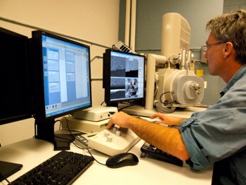
All of our instruments are networked for ease of data transfer and processing, and extensive computer hardware and software are available to support all of the instruments.
Laboratory Personnel:
Mathew Elliot: melliot@uwyo.edu
Lab Phone: 307.766.5308
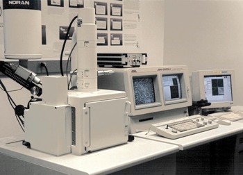
Electron Probe Microanalysis (EPMA)
Applications: This instrument is optimized for quantitative nondestructive chemical analysis of solid materials. Elements present in concentrations of at least 0.10 w% can usually be quantified to within + 1% of the measured abundance, and elements present in smaller concentrations can be measured with somewhat less precision. Any number of elements can be included in an analysis. The instrument is also capable of high-precision x-ray mapping of spatial variations in chemical compositions. Both energy dispersive spectroscopy (EDS) and wavelength dispersive spectroscopy (WDS) mapping are available.
Instrumentation: A JEOL JXA-8900 electron microprobe with five fully automated crystal spectrometers, a fully integrated EDS detector, backscattered and secondary electron detectors capable of “real-time” imaging, fully automated sample stage capable of accommodating a wide variety of sample sizes and shapes (up to six standard petrographic thin sections), and an HP workstation and peripherals for controlling data acquisition and data processing.
Sample Requirements: For optimal results, samples should be stable in a vacuum of 10-6 torr, be amenable to polishing to one micron smoothness, include no more than one element with atomic number less than five, and have individual particles of at least five microns in diameter. Smaller particles, unpolished samples, and samples with several light elements can also be analyzed using special procedures.
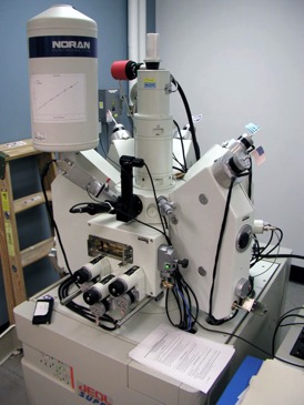
Field Emission Scanning Electron Microscopy (FESEM)
 Applications: The field emission scanning electron microscope is optimized for imaging and analysis
of shapes and spatial variations in properties such as chemical composition, crystalline
phase distributions, and analysis of orientations of individual crystals within a
polycrystalline sample. The FESEM has the highest available spatial resolution of
any instrument in our laboratories, and can resolve features on the order of one nanometer
in diameter under optimal conditions. In addition to analytical and imaging capabilities,
the FESEM is also capable of electron beam lithography, which is an important technique
in numerous fields including engineering and physics in addition to geological sciences.
Applications: The field emission scanning electron microscope is optimized for imaging and analysis
of shapes and spatial variations in properties such as chemical composition, crystalline
phase distributions, and analysis of orientations of individual crystals within a
polycrystalline sample. The FESEM has the highest available spatial resolution of
any instrument in our laboratories, and can resolve features on the order of one nanometer
in diameter under optimal conditions. In addition to analytical and imaging capabilities,
the FESEM is also capable of electron beam lithography, which is an important technique
in numerous fields including engineering and physics in addition to geological sciences.
Instrumentation: An FEI Quanta FEG 450 field emission scanning electron microscope capable of operation in high vacuum, low vacuum, and environmental modes. This instrument is equipped with a secondary electron detector, a backscattered electron detector, an Oxford Inca x-ray analysis system, an Oxford HKL diffracted backscattered electron system (EBSD), a cathodoluminesence detector (CL), an electron beam induced current detector (EBIC), and a scanning transmitted electron detector (STEM). It is also equipped with a Nabity electron beam lithography system (EBL).
Sample Requirements: Virtually any solid material can be examined in this instrument.
Conventional Scanning Electron Microscopy
 Applications: Scanning electron microscopes are optimized for imaging and analysis of shapes and
spatial variations in features such as chemical composition. The most familiar application
of SEM’s is in generating photos of extremely small details of almost any kind of
sample. Our conventional SEM is capable of spatial resolution on the order of 50 –
100 nanometers. Samples requiring higher resolution must be examined in the FESEM.
Applications: Scanning electron microscopes are optimized for imaging and analysis of shapes and
spatial variations in features such as chemical composition. The most familiar application
of SEM’s is in generating photos of extremely small details of almost any kind of
sample. Our conventional SEM is capable of spatial resolution on the order of 50 –
100 nanometers. Samples requiring higher resolution must be examined in the FESEM.
Instrumentation: We have a FEI Quanta 250 SEM equipped with a backscattered electron detector, and Oxford Inca energy dispersive xray spectroscopy system (EDS). In addition to routine high-vacuum operation, this microscope is capable of operating in a “low vacuum” mode which enables examination of samples without application of a conductive coating, and examination of samples under somewhat lower vacuum conditions.
Sample Requirements: Almost any solid material stable in a vacuum can be examined in the conventional scanning electron microscope. Biological samples, however, may require extensive sample preparation before examination in the SEM.
X-Ray Diffraction
 Applications: X-ray powder diffraction is used to examine the crystalline structure of solid materials.
It is used to identify individual phases or phases present in mixtures, to refine
crystalline structure of phases, to measure abundances of phases in polyphase mixtures
(e.g., minerals in rocks), and in some cases, to estimate sizes of extremely small
crystallites.
Applications: X-ray powder diffraction is used to examine the crystalline structure of solid materials.
It is used to identify individual phases or phases present in mixtures, to refine
crystalline structure of phases, to measure abundances of phases in polyphase mixtures
(e.g., minerals in rocks), and in some cases, to estimate sizes of extremely small
crystallites.
Instrumentation: We have a Rigaku SmartLab SE automated powder diffraction system equipped with a theta-theta goniometer and a solid state x-ray detector. The system uses SmartLab Studio II software, and is supported by extensive software for data acquisition and processing, including background subtraction, peak recognition, automated search-match procedures for phase identification, crystallography routines including peak indexing and unit cell refinements, automated quantitative analysis of mixtures, and crystallite size determination. Rietveld refinement software is also available.
Sample Requirements: Routine operation utilizes 0.05 to 0.10 grams of finely powdered sample. The sample is not damaged during analysis and can be fully recovered. This instrument has a horizontal sample stage, so liquids with suspended crystalline material can also be examined. Coarse-grained and single-crystal samples can also be examined using special techniques.
X-Ray Fluorescence Analysis
Applications: This instrument is used for quantitative analysis of the bulk chemistry of solid and in some cases liquid samples. Soils and rocks are the primary types of samples analyzed in our lab, but any solid can in principle be analyzed. Major elements can usually be measured down to 0.02-0.05 w%; the accuracy usually is ±1% of the measured concentration for higher abundance elements, and somewhat less for lower abundance elements. Trace elements can be measured down to concentrations of 2-20 ppm depending on the element.
Instrumentation: Our instrument is a PANalytical Axios XRF Analyzer equipped with a 4000W Rh SST-max x-ray tube and three wavelength-dispersive spectrometers. The instrument includes a robotic sample changer which allows us to run batches of up to 96 samples in an automated mode. Instrument control and data acquisition and processing are performed on a Windows workstation. We have three software packages for different types of analyses. The basic IQ+ analysis package is a general application for the quantitative analysis of all elements from O to U and is used primarily for samples of unknown chemical composition or fast semi-quantitative analysis of irregularly shaped samples. The WROXI (wide-range oxide analyses) package is specifically designed and calibrated for oxide analyses in rocks and soils. Elements included in this set are Si, Al, Na, K, Mg, Ca, V, Cr, Ni, Cu, Ti, Fe, Mn, Zn, Zr, Sr, Ba, Pb, Hf, S and P. Trace elements are analyzed using the ProTrace package, which allows for the analysis of Sc, V, Cr, Co, Ni, Cu, Zn, Ga, Ge, As, Se, Br, Rb, Sr, Y, Zr, Nb, Mo, Ag, Cd, Sn, Sb, Te, I, Cs, Ba, La, Ce, Nd, Sm, Yb, Hf, Ta, W, Tl, Pb, Bi, Th and U.
Sample Requirements: For most accurate results, samples should be finely ground and either fused into a glass bead for the analysis of major elements or pressed into a solid pellet for trace element analyses. For a complete analysis, both a fused glass bead and a pressed pellet are commonly prepared. Approximately 1 g of sample is needed for glass bead preparation, and 4 g for producing a pressed pellet. While these preparation methods are destructive, samples can also be measured non-destructively as loose powders or solid pieces, although accuracy will be lower for these measurements for a number of reasons. Accuracy can be improved if the sample is as homogeneous as possible and if solid pieces have polished or at least reasonably flat surfaces. Our sample cups can handle objects between roughly 10 mm and 30 mm in size.
The University of Wyoming has earned its Research Level 1 (R1) status from the Carnegie Classification of Institutions of Higher Education, placing Wyoming's only four-year university with the top research universities in the United States.