This is a differential medium. It is a rich, complex medium that contains 5% sheep red blood cells. BAP tests the ability of an organism to produce hemolysins, enzymes that damage/lyse red blood cells (erythrocytes). The degree of hemolysis by these hemolysins is helpful in differentiating members of the genera Staphylococcus, Streptococcus and Enterococcus.
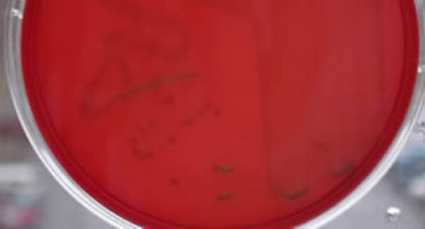
Beta-hemolysis is complete hemolysis. It is characterized by a clear (transparent) zone surrounding the colonies. Staphylococcus aureus, Streptococcus pyogenes and Streptococcus agalactiaeare b-hemolytic (the picture on the right below shows the beta-hemolysis of pyogenes).
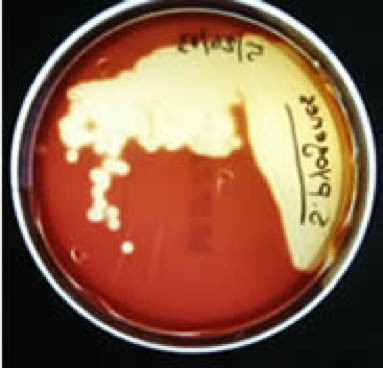
Partial hemolysis is termed alpha-hemolysis. Colonies typically are surrounded by a green, opaque zone. Streptococcus pneumoniaeand Streptococcus mitis are a-hemolytic (the picture in the middle below shows the a-hemolysis of S. mitis).
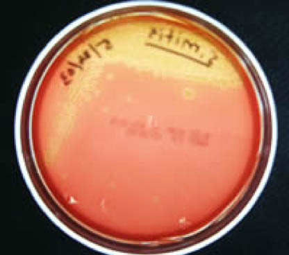
If no hemolysis occurs, this is termed gamma-hemolysis. There are no notable zones around the colonies. Staphylococcus epidermidisis gamma-hemolytic.
 |
 |
 |
| Gamma-hemolysis | Alpha-hemolysis | Beta-hemolysis |
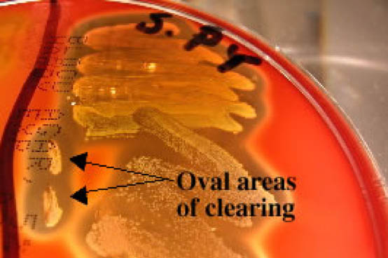
Often when inoculating a BAP to observe hemoloysis patterns, investigators will also stab several times through the agar using an inoculating loop. This stab allows for the detection of streptolysin O, a specific hemolysin produced by Streptococcus pyogenes. This hemolysin is inactivated by O2 and is only seen subsurface (in an anaerobic environment) around the stab mark. Note the oval-shaped areas of clearing around the stab marks in the picture below; these are caused by streptolysin O.