CASI prioritizes University of Wyoming researchers, providing them with specialized services in microscope imaging and X-ray analyses. Our commitment extends to supporting and training faculty and student researchers across campus. While prioritizing UW researchers, CASI also extends its services to regional and nationwide contract users, ensuring broad accessibility to our state-of-the-art facilities. Additional information is available in the Resources and Training and Proposals sections.
Please explore the descriptions provided for comprehensive details about our instruments, including the instrument manager, location, fees, specifications, training requirements, and reservation options. To access more in-depth information about each instrument, click on the instrument image or name, directing you to dedicated pages. Additionally, refer to the Rates page for specific fee details.
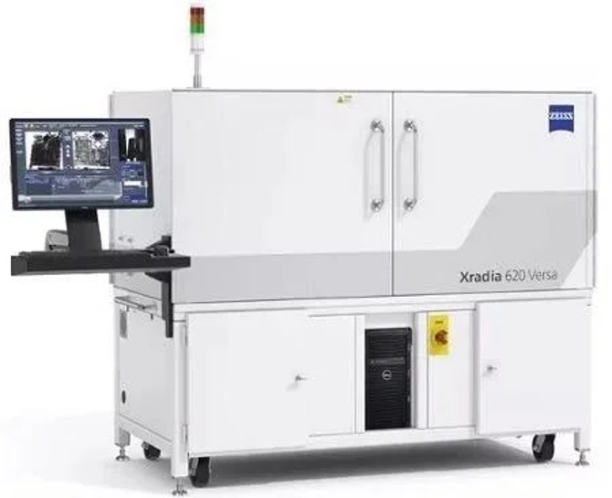
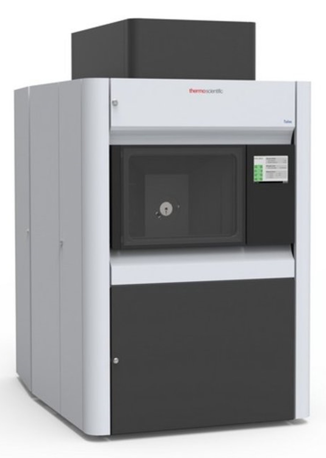
Allows for high resolution, detailed, non-destructive x-ray scans of the internal anatomy of animals and museum species without any damage to the specimens, giving important insight into the functioning of artifacts. The Micro-CT can be used in applications related to organismal biology, engineering and materials science, biomedical imaging, archaeology and paleontology, and geology.
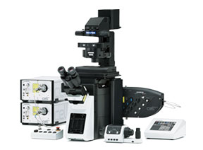
Generates a confocal image using a spinning, opaque disk with hundreds of pinholes arranged in spirals, rotating at high speeds, to scan across a sample. The microscope then images multiple, thin 2D slices of a sample and constructs a 3D model from them. The microscope can study fast dynamic processes, long-term time-lapses, or details inside the cell membrane, all possible with live cells, and can be used across a wide range of biological sciences. This system is also equipped with TIRF functionality to explore a very thin optical section close to the coverslip with high signal to noise.
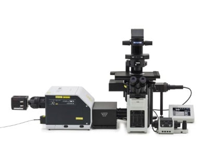
"spINBRE SuperRes": SD Confocal
Generates a confocal image using a spinning, opaque disk with hundreds of pinholes arranged in spirals, rotating at high speeds, to scan across a sample. The microscope can be used to image multiple, thin 2D slices of a sample and constructs a 3D model from them. The microscope can study fast dynamic processes, long-term time-lapses, or details inside the cell membrane, all possible with live cells, and can be used across a wide range of biological sciences. This system is equipped with super-resolution functionality to get resolutions below the diffraction limit.
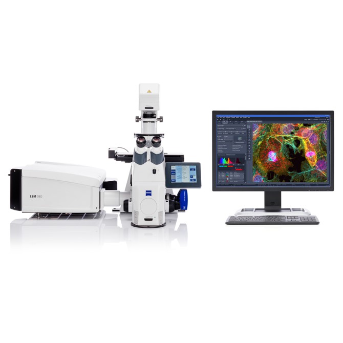
Laser-scanning confocal microscope for multiplex and NIR imaging
Images atomic scale structure using a beam of electrons passing through an ultrathin specimens. Applications are wide-ranging from life sciences, nanotechnology, medical research, and materials science.
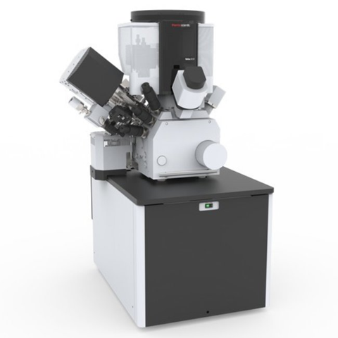
Images atomic scale structure using a focused beam of ions and allows scientists to generate 3D reconstructions and to mill and manufacture nano structures. Applications are wide-ranging from agriculture, life sciences, microbiology, earth science, the oil and gas industry, battery and solar cell research, and materials science.
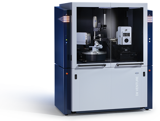
"Old Faithful": Single Crystal XRD
Provides detailed information about the internal lattice of crystalline molecular solids, including unit cell dimensions, bond-lengths, bond-angles, and details of site-ordering. Applications range from chemistry, molecular biology, pharmacy, physics, and chemical engineering.