
Grasshoppers of Wyoming and the West
Entomology
Detailed External Anatomy
Head
The head of the grasshopper is a hard capsule that contains large muscles, which operate the chewing mouthparts, and the brain and subesophageal ganglion, which serve as the main centers of the nervous system. Prominent on the outside of the capsule are a pair of antennae, two large compound eyes, and the downward directed mouthparts. The antennae of grasshoppers are usually filiform (threadlike) but they may have other shapes, such as ensiform (broad at base, narrowing to tip) or clavate (expanded at tip) (Fig. 2). Compound eyes vary in shape and protuberance. They are usually somewhat round but may be elliptical in grasshoppers with strongly slanted faces.
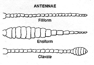 |
Figure 2. Diagram of three forms of grasshopper antennae: filiform or threadlike, ensiform or sword-shaped, and clavate or club shaped |
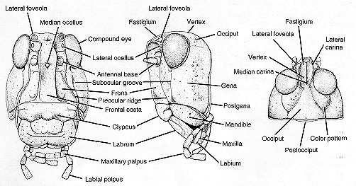 |
Figure 3. Grasshopper head, front, side, and top views. Modeled after Trimerotropis pallidipennis (Burmeister). |
The head capsule is divided into areas by visible sutures, external ridges (carinae), or by general location (Fig. 3). The top of the head between the compound eyes is known as the vertex. Behind the vertex is the occiput, and in front of the vertex is the fastigium. A pair of variously shaped depressions, the lateral foveolae, is often present in front or at the sides of the fastigium. The front of the head between the compound eyes and extending to the clypeus is known as the frons. A wide ridge, the frontal costa, runs down the middle of the frons from the fastigium toward the margin of the clypeus. The side of the head below the compound eye is named the gena or cheek. Grasshoppers have three simple eyes called ocelli — one above the base of each antenna and one centrally located in the frontal costa. These and other parts and appendages of the head are illustrated in Figure 3.
Thorax
The thorax, locomotion center of the grass-hopper, is a stout, boxlike structure consisting of three fused segments: the prothorax, mesothorax, and metathorax. Each segment bears a pair of legs. The second segment bears a pair of fore-wings, the tegmina, and the third segment a pair of membranous hindwings. The wings of a few species are reduced to small pads or are entirely lacking. The top of the thoracic segments is called the notum, the bottom the sternum, and the sides the pleura.
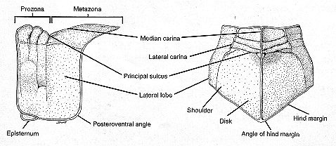 |
Figure 4. Grasshopper pronotum, side and top views. Modeled after Trimerotropis pallidpennis (Burmeister). |
The pronotum situated just behind the head is a prominent, saddle-shaped structure with lateral lobes that hide nearly all of the propleura (Fig. 4). The pronotum has many distinctive features useful in separating both genera and species of grasshoppers. The integument (skin) may be nearly smooth in some species and rough and wrinkled in others. The dorsum or disk of the pronotum is divided into left and right halves by a longitudinal ridge, the median carina. The ridge varies among species from barely visible to a conspicuously high crest. Transverse furrows run across the disk and down the lateral lobes. These furrows, known as sulci, cut into the median carina and divide the disk into zones, the prozona in front and the metazona in the rear. In many species only one sulcus cuts the median carina while in others two or three sulci cut the median carina. The hind sulcus is considered the principal sulcus; from its position the length of the prozona and metazona are measured.
The lateral lobes usually form an angle with the disk and are separated from the disk by lateral carinae that, depending on the species, may be straight and parallel or variously incurved or outcurved. The hind margin of the disk varies from an acute angle to an obtuse angle, or may be convex, truncate, or emarginate.
The various shapes, sizes, and protuberance of the sternal sclerites afford reliable taxonomic characters (Fig. 5). A prosternal spine located between the bases of the front legs is characteristic of members of the spurthroated subfamily. Shapes and dimensions of the mesosternal and metasternal lobes and interspaces are useful in separating certain species and subfamilies.
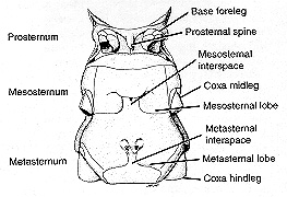 |
Figure 5. Sternum of thorax, bottom view. Modeled after Melanoplus bivittatus (Say) female. |
Legs
Although the three pairs of legs have the same component parts, the hind pair, adapted for jumping, are much larger than the first and second pair and bear more distinctive features. The color and markings of both the femur and tibia differ among species. The robust femur has several surfaces and ridges that have been given names for easy reference (Fig. 6).
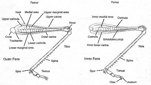 |
Figure 6. Grasshopper hindleg, views of outer and inner faces. Hindleg of Mermiria bivittata (Serville). |
The long and slender tibia bears along its posterior edges a double row of spines and distally two pairs of articulated spurs or calcars. The number of spines and the length of calcars vary among species. The inner medial area of the femur may have a longitudinal ridge bearing a series of stridulatory pegs. Up and down movements of the hindlegs cause the pegs to scrape against a raised vein on each tegmen, which produces a song or signal peculiar to that species of grasshopper.
Wings
The two pairs of grasshopper wings differ in shape, structure, and function (Fig. 7). The front pair, or tegmina, are leathery and narrow with the sides nearly parallel. The hind wings are membranous and fan-shaped. Compared with the tegmina, the hind pair contribute three times as much to flight lift. Both pairs afford diagnostic characters that aid in the identification of species. The wing veins, sclerotized tubes providing strength to the wings, vary greatly in thickness. The tegmina vary from immaculate to distinctly spotted or marked. The hindwings of grasshoppers are usually hyaline. Members of one subfamily, the Oedipodinae or bandwinged grasshoppers, have wings with a dark submarginal band and have the disk colored.
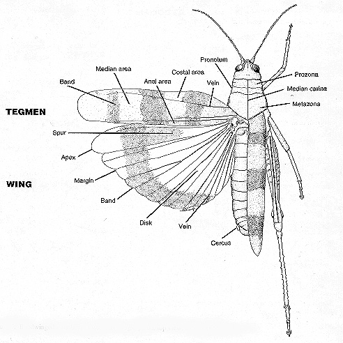 |
Figure 7. Bandwinged grasshopper with left wings spread, top view. Composite model |
Abdomen
The hind region of the grasshopper’s body, the abdomen, consists of 11 segments (Fig. 1). Segment I is firmly fused with the metathorax and contains the auditory organ with its eardrum cover, the tympanum (Fig. 8).
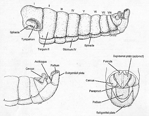 |
Figure 8. Grasshopper male abdomen, side view and enlarged side and dorsal views of end. Modeled after Melanoplus packardii Scudder. |
Segments II to VIII are ringlike in appearance and are separated from one another by pliable membranes. Each segment has a sclerotized tergum that covers not only the top but also the sides of the abdomen. A sclerotized sternum covers the bottom. Pliable membranes separate the terga from the sterna and with the intersegmental membranes allow the abdomen much flexibility, a requirement for respiratory movements, copulation, and oviposition.
Genitalia
The terminal segments of the abdomen are reduced and modified to bear the external reproductive organs, the genitalia, and the associated structures (Fig. 8). These structures offer the most reliable taxonomic characters for separating spurthroated grasshoppers. Structures of the male are more distinctive than those of the female. The prominent paired cerci are usually conical, but in the males of some genera, e.g. Melanoplus, they have characteristic sizes and shapes. Likewise, the furcula, a pair of projections from the posterior edge of tergum X of males, differs in size and shape. The epiproct or supraanal plate, although roughly triangular, varies sufficiently in shape and rugosity to be taxonomically useful. The variations in shape and protuberances of the subgenital plate are also useful in identification. These structures are easily seen with a pocket magnifier of 10x magnification. A few distinctive structures, such as the lobes of the aedeagus, require the use of a stereomicroscope (magnification of 50x and greater) for clear identification.
The valves of the ovipositor are sometimes useful in separating species (Fig 1). The dorsal and ventral pair of valves have various shapes and denticulations. The middle pair of valves are small and hidden.
The sclerotized integument of the abdomen varies in color, patterns, and texture among species and sometimes affords distinguishing taxonomic characters.