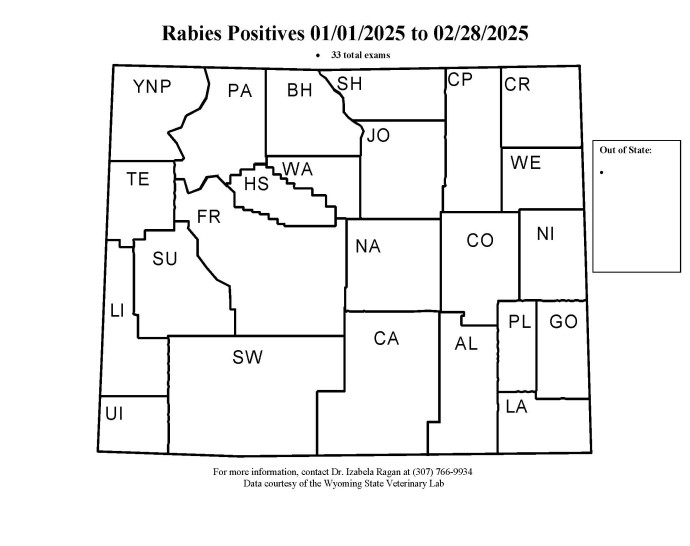
Rabies Transmission and Exposure Explained
Spread of rabies commonly occurs through a bite from an infected animal. Once an animal or human has been exposed to the rabies virus, the virus travels from the peripheral nervous system (PNS) to the central nervous system (CNS) to increase replication, and then travels back through the PNS, which includes the salivary glands. The infected saliva is the medium that transmits the virus when a bite occurs. While bites are the most common route of transmission, any exposure to infected bodily fluids through cuts or abrasions in the skin can allow the virus to infect a new host as well. Inhalation of virus-containing aerosols, contacting raw meat products of an infected animal, and through organ transplant transmission has been reported but is extremely rare.
Depending on the severity of exposure, post-exposure prophylaxis (PEP) may be needed. Contact your primary physician and/or your local public health department to assess exposure risk and determine whether PEP is warranted in human exposure cases. According to the World Health Organization (WHO), exposure to a suspect rabid animal is defined as:
Category I: Touching or feeding animals, animal licks intact skin (no exposure)
Category II: Scratches or abrasions with no bleeding that were obtained from the animal (exposure)
Category III: Transdermal bites, contamination of mucous membranes or broken skin with saliva from
the animal, direct contact with brain/spinal cord tissue, contact with bats (severe
exposure)
Testing for Rabies at WSVL
Rabies testing in animals is essential for confirming diagnosis, tracking disease spread, and protecting public health. Testing at the WSVL is performed postmortem because our assay requires testing of brain tissue, where the virus concentrates.
All in-state testing for rabies is free of charge. WSVL will add a $50.00 charge to all out-of-state rabies submissions. Please contact the lab at vetrec@uwyo.edu with questions.
For information on how to properly ship us your samples, please click below.
Rabies Sample Collection Flyer Packing and Shipping Samples
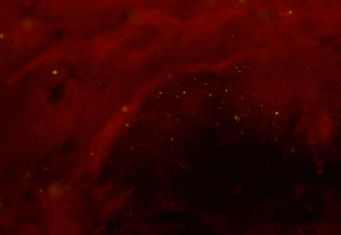
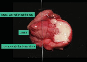
At the WSVL we perform the Direct Fluorescent Antibody (DFA) test, currently the gold standard test for rabies diagnosis in animals. To perform the test, a complete cross-section is made through the cerebellum and brainstem, then impression slides are made from these tissues. These slides are fixed and incubated with fluorescently-labeled antibodies. Unbound antibodies are washed away in the final processing step, but areas where rabies antigen are present, the antibodies are bound. These antibodies can be visualized as a fluorescent-apple-green color using a fluorescence microscope.
Positive
When rabies antigen is present within a sample, the fluorescently-labeled antibodies appear green under the fluorescence microscope.

Negative
When rabies antigen is absent from a sample, the fluorescently-labeled antibodies are washed away in the final rinse step, and no green fluorescence is observed.
Suitable Sample Guide
Images obtained from the "Protocol for Postmortem Diagnosis of Rabies in Animals by
Direct
Fluorescent Antibody Testing" provided by the Centers for Disease Control and Prevention
(CDC)
Unsuitable Samples
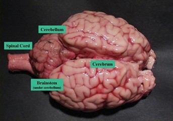
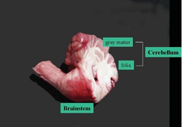
Brainstem is required for diagnostic testing. A strict surveillance sample, with no human or animal exposures, may be diagnosed with only the brainstem per the Centers for Disease Control (CDC). However, if there is any known or even suspected exposure to humans or animals, a complete cross-section of the brainstem and cerebellum are required. Cross-sections of both sides of the hippocampus may be substituted if no cerebellum is present. If the required samples are not received, not identifiable, or autolyzed, the diagnosis will be listed as “unsuitable” unless the rabies antigen is detected, which would indicate a positive case. If no fluorescence is observed, and the sample is reported as “unsuitable,” it simply means the sample we were provided is not suitable for diagnosis; rabies cannot be ruled out. If no brain material is submitted and rabies testing is requested, the submitter will be contacted and no testing will be performed. Samples that are initially reported as "Inconclusive" by DFA will be confirmed by a secondary test.
Bats
Bats are a primary reservoir globally. They are an important reservoir in Wyoming.
Image Credit: Healthy little brown bats by Ann Froschauer , Ann Froschauer /USFWS , Public Domain, https://www.fws.gov/media/healthy-little-brown-bats-ann-froschauer
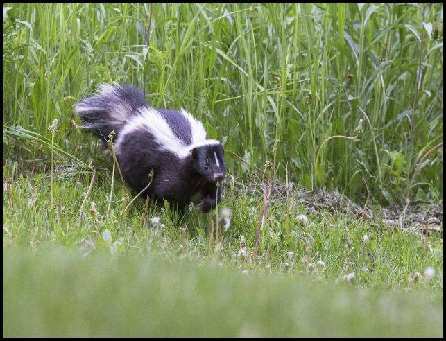
Skunks
A major terrestrial reservoirs in North America. Skunks are an important reservoir in Wyoming.
Image Credit: Striped Skunk, K Theule/USFWS, Public Domain, https://www.fws.gov/media/striped-skunk-0
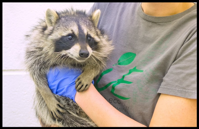
Raccoons and Foxes
Both foxes and raccoons are major reservoirs in North America.
Image Credit: Raccoon on the move, Burton, Robert H./USFWS, Public Domain, https://www.fws.gov/media/raccoon-move and Lone red fox standing amongst rocks, Keepers, Holly/USFWS, Public Domain, https://www.fws.gov/media/lone-red-fox-standing-amongst-rocks
Dogs and Cats
Dogs and cats can become infected. Dogs are responsible for a significant number of human rabies deaths in developing countries. Routine rabies vaccination is highly effective and remains the best way to protect dogs and cats.
In Wyoming, rabies is most commonly identified in wildlife species such as bats, skunks, and occasionally other wild carnivores. Domestic animals, including cats, dogs, horses, and livestock, can become infected following exposure to rabid wildlife, creating a risk for both animal and human health.
Informational Rabies links to various external agencies are available below for further reading:
Centers for Disease Control (CDC)
United States Department of Agriculture (USDA)
World Health Organization (WHO)