
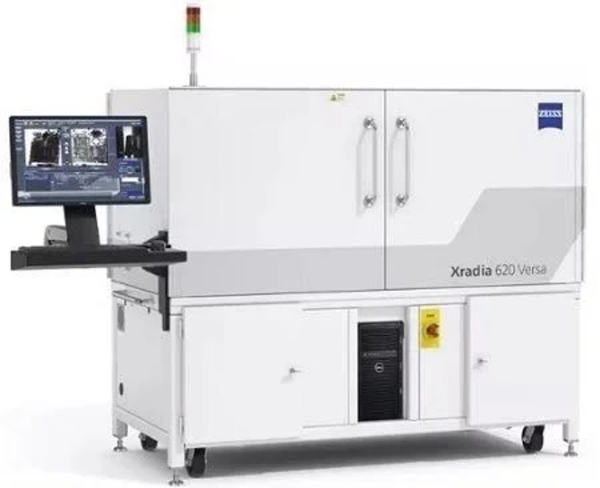
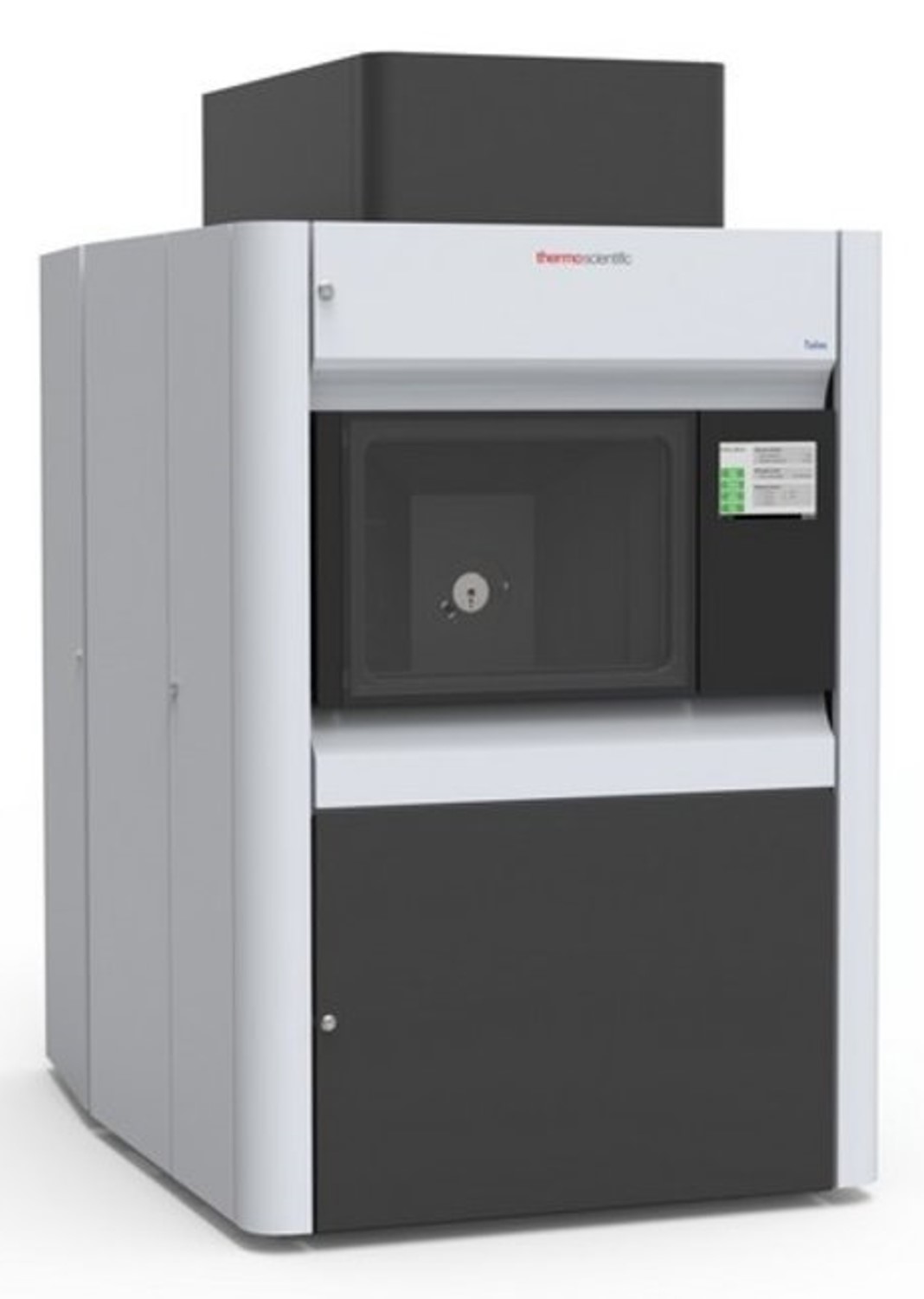
Allows for high resolution, detailed, non-destructive x-ray scans of the internal anatomy of animals and museum species without any damage to the specimens, giving important insight into the functioning of artifacts. The Micro-CT can be used in applications related to organismal biology, engineering and materials science, biomedical imaging, archaeology and paleontology, and geology.
- Zeiss Xradia 610 Versa
- Instrument Manager: Qian Yang, qyang3@uwyo.edu
- Instrument Location: Science Initiative Building Bay 4, 1305
- Fees are listed in Rates
- To book this instrument and request training please go to the PPMS login page
- Use of this instrument requires mandatory training sessions
- Service can be provided
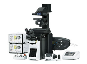
Generates a confocal image using a spinning, opaque disk with hundreds of pinholes arranged in spirals, rotating at high speeds, to scan across a sample. The microscope then images multiple, thin 2D slices of a sample and constructs a 3D model from them. The microscope can study fast dynamic processes, long-term time-lapses, or details inside the cell membrane, all possible with live cells, and can be used across a wide range of biological sciences. This system is also equipped with TIRF functionality to explore a very thin optical section close to the coverslip with high signal to noise.
- Olympus IXplore
- Instrument Manager:
- Instrument Location: Science Initiative Building Bay 1, 1305
- Fees are listed in Rates
- To book this instrument and request training please go to the PPMS login page
- Use of this instrument requires mandatory training sessions
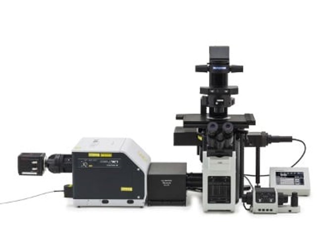
"spINBRE SuperRes": SD Confocal
Generates a confocal image using a spinning, opaque disk with hundreds of pinholes arranged in spirals, rotating at high speeds, to scan across a sample. The microscope can be used to image multiple, thin 2D slices of a sample and constructs a 3D model from them. The microscope can study fast dynamic processes, long-term time-lapses, or details inside the cell membrane, all possible with live cells, and can be used across a wide range of biological sciences. This system is equipped with super-resolution functionality to get resolutions below the diffraction limit.
- Olympus IXplore
- Instrument Manager:
- Instrument Location: Science Initiative Building Bay 2, 1305
- Fees are listed in Rates
- To book this instrument and request training please go to the PPMS login page
- Use of this instrument requires mandatory training sessions
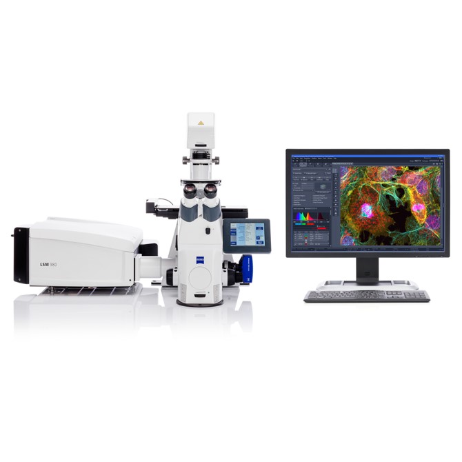
Laser-scanning confocal microscope for multiplex and NIR imaging
- Zeiss LSM 980 w/ AiryScan 2
- Instrument Manager:
- Instrument Location: Science Initiative Building Bay 3, 1305
- Fees are listed in Rates
- To book this instrument and request training please go to the PPMS login page
- Use of this instrument requires mandatory training sessions
Images atomic scale structure using a beam of electrons passing through an ultrathin specimens. Applications are wide-ranging from life sciences, nanotechnology, medical research, and materials science.
- ThermoFisher Talos F200X G2
- Instrument Manager: Qian Yang, qyang3@uwyo.edu Science Initiative Building 1311
- Fees are listed in Rates
- To book this instrument and request training please go to the PPMS login page
- Use of this instrument requires mandatory training sessions
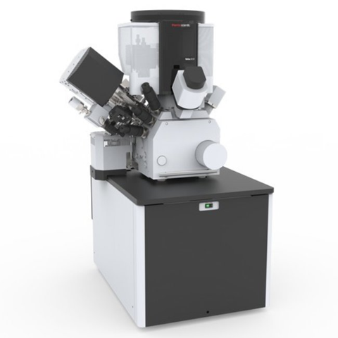
Images atomic scale structure using a focused beam of ions and allows scientists to generate 3D reconstructions and to mill and manufacture nano structures. Applications are wide-ranging from agriculture, life sciences, microbiology, earth science, the oil and gas industry, battery and solar cell research, and materials science.
- ThermoFisher Helios 5 UX
- Instrument Manager: Qian Yang, qyang3@uwyo.edu
- Science Initiative Building 1315
- Fees are listed in Rates
- To book this instrument and request training please go to the PPMS login page
- Use of this instrument requires mandatory training sessions
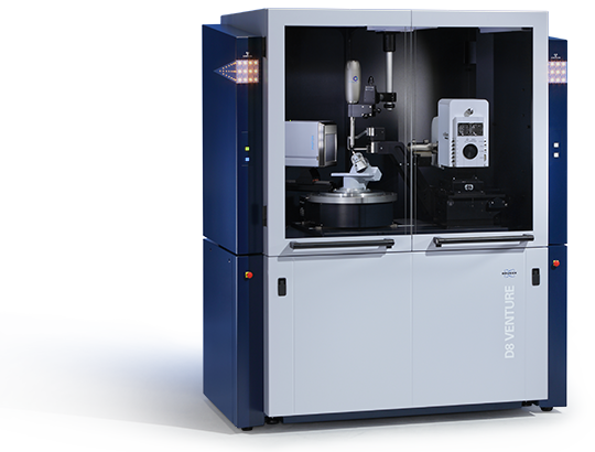
"Old Faithful": Single Crystal XRD
Provides detailed information about the internal lattice of crystalline molecular solids, including unit cell dimensions, bond-lengths, bond-angles, and details of site-ordering. Applications range from chemistry, molecular biology, pharmacy, physics, and chemical engineering.
- Bruker D8 Venture
- Instrument Manager: Qian Yang, qyang3@uwyo.edu
- Science Initiative Building Bay 1, Showcase 3010
- Fees are listed in Rates
- To book this instrument and request training please go to the PPMS login page
- Use of this instrument requires mandatory training sessions
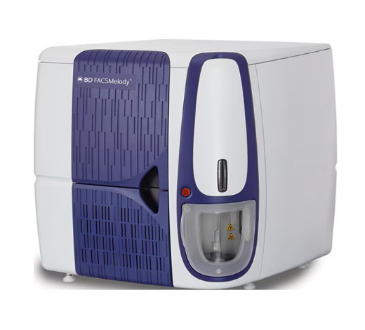
The "Snake River": Cell Sorter
BD FACSMelody™ Cell Sorter is a flow cytometry system designed to simplify cell sorting for researchers. Featuring advanced automation through BD FACSChorus™ software, it guides users with an intuitive interface, on-screen instructions, and detailed reports. With up to 11 parameters and 4-way sorting capability, it supports a wide range of applications, delivering high sensitivity and resolution to isolate target cells efficiently. Its streamlined workflow, quick setup, and fixed alignment technology minimize hands-on time, making it an accessible, cost-effective solution for in-house cell sorting.
- Instrument Manager: Steven Florez
- Engineering Education and Research Building (EERB) 115
- To book this instrument and request training please go to the PPMS login page
- Use of this instrument requires mandatory training sessions
The University of Wyoming has earned its Research Level 1 (R1) status from the Carnegie Classification of Institutions of Higher Education, placing Wyoming's only four-year university with the top research universities in the United States.